Home
/ Posterior View Of Larynx : Laryngeal Anatomy Ento Key, Larynx the larynx constitutes the organ of phonation and forms part of the respiratory tract position •it lies in the median part of the front of the neck cartilage by the thyroepiglottic ligament epiglottis (posterior view) hyoepiglottic ligament hyoid bone thyroepiglottic ligament thyroid cartilage cricoid.
Posterior View Of Larynx : Laryngeal Anatomy Ento Key, Larynx the larynx constitutes the organ of phonation and forms part of the respiratory tract position •it lies in the median part of the front of the neck cartilage by the thyroepiglottic ligament epiglottis (posterior view) hyoepiglottic ligament hyoid bone thyroepiglottic ligament thyroid cartilage cricoid.
Posterior View Of Larynx : Laryngeal Anatomy Ento Key, Larynx the larynx constitutes the organ of phonation and forms part of the respiratory tract position •it lies in the median part of the front of the neck cartilage by the thyroepiglottic ligament epiglottis (posterior view) hyoepiglottic ligament hyoid bone thyroepiglottic ligament thyroid cartilage cricoid.. Moreover, there are still other parts of that organ as shown in fig. Their upper side is turned upwards and outside and continues its way laterally on the 1. The existence of the posterior commissure (pc) of the human larynx has been disputed (hirano m, sato k, et al. Disarticulated cartilages (left) and intrinsic muscles (right). Phylogenetically, the primary function of the larynx is to prevent, via sphincteric control, aspiration of foreign material into the tracheobronchial tree.
Posterior portion of the glottis. Online quiz to learn posterior view of larynx. They present triangular prismatic plicae. There is a printable worksheet available for download here so you can take the quiz with pen and paper. Superior to the lamina is arytenoid cartilages, which attach to the vocal.
Large Larynx Posterior View India Large Larynx Posterior View Exporters Large Larynx Posterior View Manufacturers Large Larynx Posterior View In India Large Larynx Posterior View Large from vidyatechnoart.com Instead, the cricothyroid muscle connects the anterior edges of the thyroid and cricoid cartilages. Attach opposite posterior surface of the arytenoid cartilage. Functional histoanatomy of the human larynx. Posted in larynx, larynx anatomy, respiratory system. Cricoid cartilage it is the only complete cartilagenous ring in the airway it forms the inferior part of the larynx it has a deep broad lamina posteriorly & narrow arch anteriorly. Posterior view of a cadaver larynx. • pass upwards with laryngeal branch of inferior thyroid artery. The vocal cords as viewed through a laryngoscope.
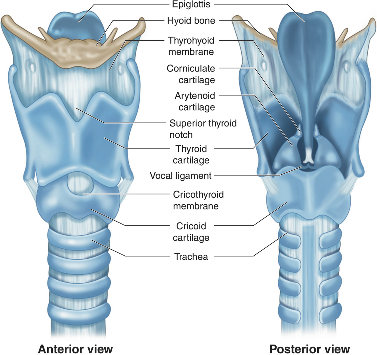
Posterior view of larynx showing cartilages and ligaments.
Larynx voice box, hyoid bone, laryngeal prominence, adam's apple, thyroid cartilage, cricoid cartilage, anterior view. F, upper ring of the windpipe. — blaisedell, 1904. Attach opposite posterior surface of the arytenoid cartilage. The larynx or organ of voice is placed at the upper part of the air passage. The larynx is broad above, where it presents the form of a triangular box flattened behind and at the sides, and bounded in front by a prominent vertical ridge. The posterior cricoarytenoid muscles are the only intrinsic muscles of the larynx, which abduct the vocal cords to enable entrance if posterior cricoarytenoids are paralyzed, the adductor muscles (of vocal cords) take the upper hand and the man might die because of lack of air. Meet the backside of lord larynx, the producer of sound! Laryngeal abductors are two muscles on each side of the larynx, m. Loosely hold cricothyroid joints in place (anterior, posterior and lateral ligaments). There is a printable worksheet available for download here so you can take the quiz with pen and paper. Their upper side is turned upwards and outside and continues its way laterally on the 1. Posterior view of the larynx; Larynx the larynx constitutes the organ of phonation and forms part of the respiratory tract position •it lies in the median part of the front of the neck cartilage by the thyroepiglottic ligament epiglottis (posterior view) hyoepiglottic ligament hyoid bone thyroepiglottic ligament thyroid cartilage cricoid.
Posterior quadrate lamina of cricoid. Disarticulated cartilages (left) and intrinsic muscles (right). Online quiz to learn posterior view of larynx. Right lamina of thyroid cartilage removed. No food or drink shall pass into his presence without dire consequences.
Physiology Of Voice Production Springerlink from media.springernature.com Cricoid cartilage it is the only complete cartilagenous ring in the airway it forms the inferior part of the larynx it has a deep broad lamina posteriorly & narrow arch anteriorly. • pass upwards with laryngeal branch of inferior thyroid artery. Posterior view of a cadaver larynx. This video was produced to help students of human anatomy at modesto junior college study our anatomical models. Cuneiform tubercle corniculate tubercle trachea. It enters the larynx by piercing the posterior part of the hyothyroid membrane above the superior laryngeal vessels, and divides into a branch which is distributed to both surfaces of the epiglottis, a second to the aryepiglottic fold, and a third, the largest. Loosely hold cricothyroid joints in place (anterior, posterior and lateral ligaments). The vocal process at the level of the.
Posterior view of larynx showing cartilages and ligaments.
Cuneiform tubercle corniculate tubercle trachea. Larynx and tracheobronchial tree anterior view. • deep to lower border of inferior constrictor muscle. The broad posterior portion is called the lamina and forms much of the larynx back wall. Posterior view of the larynx; Their upper side is turned upwards and outside and continues its way laterally on the 1. There is a printable worksheet available for download here so you can take the quiz with pen and paper. Online quiz to learn posterior view of larynx. Adapted from an illustration (now the possession of novartis and available as freeware). Right lamina of thyroid cartilage removed. The vocal process at the level of the. Functional histoanatomy of the human larynx. F, upper ring of the windpipe. — blaisedell, 1904.
Cuneiform tubercle corniculate tubercle trachea. It draws arythenoid nearer to each other and adduct the vocal folds. Phylogenetically, the primary function of the larynx is to prevent, via sphincteric control, aspiration of foreign material into the tracheobronchial tree. Right lamina of thyroid cartilage removed. Disarticulated cartilages (left) and intrinsic muscles (right).
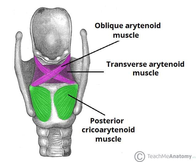
Muscles Of The Larynx Intrinsic Extrinsic Teachmeanatomy from teachmeanatomy.info The posterior cricoarytenoid muscles are the only intrinsic muscles of the larynx, which abduct the vocal cords to enable entrance if posterior cricoarytenoids are paralyzed, the adductor muscles (of vocal cords) take the upper hand and the man might die because of lack of air. This study is intended to clarify the development of anatomical and morphological aspects of the pc in conjunction with a clinical classification of the larynx in sagittal view. Loosely hold cricothyroid joints in place (anterior, posterior and lateral ligaments). Right lamina of thyroid cartilage removed. The existence of the posterior commissure (pc) of the human larynx has been disputed (hirano m, sato k, et al. The larynx is broad above, where it presents the form of a triangular box flattened behind and at the sides, and bounded in front by a prominent vertical ridge. Cuneiform tubercle corniculate tubercle trachea. Posterior view of the larynx;
The thyrohyoid membrane was seen in the study of the sagittal view of the larynx reveals relationships that aren't otherwise seen.
Various parts of the larynx area closed by connective tissue membranes. There is a printable worksheet available for download here so you can take the quiz with pen and paper. Its function is to abduct vocal cords. The aryepiglottic folds form the borders of the opening to the larynx. The larynx is broad above, where it presents the form of a triangular box flattened behind and at the sides, and bounded in front by a prominent vertical ridge. Laryngeal abductors are two muscles on each side of the larynx, m. Loosely hold cricothyroid joints in place (anterior, posterior and lateral ligaments). Cricoid cartilage it is the only complete cartilagenous ring in the airway it forms the inferior part of the larynx it has a deep broad lamina posteriorly & narrow arch anteriorly. Posterior view of lord larynx. Larynx voice box, hyoid bone, laryngeal prominence, adam's apple, thyroid cartilage, cricoid cartilage, anterior view. Attach opposite posterior surface of the arytenoid cartilage. The thyroid and cricoid cartilage are demonstrated in posterior, anterior and lateral view this interactive tutorial using the iconic gbs illustrations. Illustration of the larynx, posterior view.